

MicroMeshes
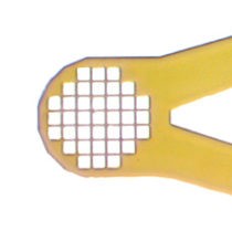
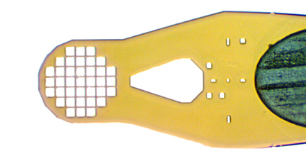
MicroMeshes™ are ideal for very small (<30 μm) samples, allowing them to be sieved out of solution. They also provide continuous and delicate support for thin plates, rods, and tissue samples.
Each box contains twenty (20) MicroMeshes™
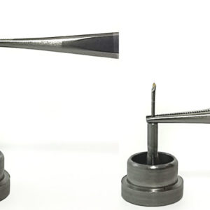
MiTeGen offers ready-to-use assemblies. Mounts can either be pre-inserted into reusable bases or epoxied into standard bases.
Reusable Mount/Base Assemblies Epoxied Mount/Base Assemblies
Product Information
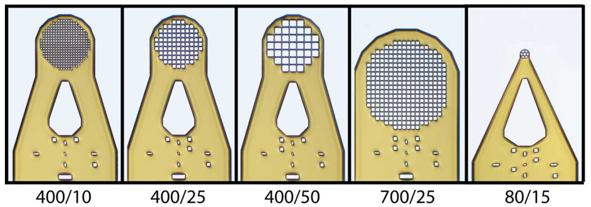
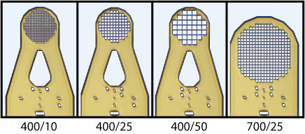
Standard MicroMeshes™ – mesh filled apertures with diameters of 400 or 700 μm and mesh openings of 10, 25 and 50 μm.
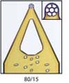
MicroMeshes SH™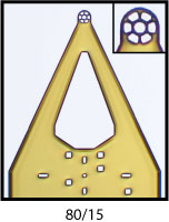 – 80 μm diameter mesh area with 15 μm openings. The small head fits entirely within the X-ray beam diameter typical of standard synchrotron beam lines and of focused laboratory sources. You don't have to see your tiny sample to align it: just center the 80 μm head in the beam, and your sample will be within it. The smaller head also makes it easier to mount a single sample, and produces less background scatter when the beam is aligned along the plane of the film.
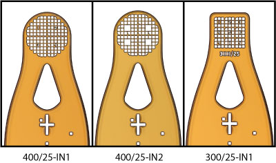
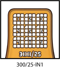
Indexed MicroMeshes™ - 300 or 400 μm mesh areas with 25 μm openings. Index marks and numbers make it easier to locate (and then to relocate) a given sample. Identify the most promising samples on your home microscope, and then easily find them again, e.g., at a microfocus X-ray source. Additional diagonal tabs allow the front/back orientation of the mesh and a crystal's position on the mesh to be uniquely determined in only a 100 μm field of view. Crystals can sometimes be more clearly seen in the larger square openings of the 400/25-IN2 design. The flat top of the 300/25-IN1 design makes it easier to scrape/scoop samples off of the bottom of a well or slide.
These styles are available in single size boxes of 20 MicroMeshes™ or as an assortment:
A1: 20 pins, 5 each of 400/10, 400/25, 400/50, and 700/25 μm head diameter/mesh aperture.
MicroMeshes™ are built on 18 mm / SPINE length rods (pins), which are now standard at nearly all synchrotron beam lines in the world. Other pin lengths are also available: (11mm, 19mm, and 25mm)
We strongly recommend using heavy-duty, serrated-tip tweezers to handle MicroMeshes™.
MicroMeshes™ are compatible with all standard X-ray hardware and can be inserted in 0.7 mm mechanical pencils or micromanipulators for easy handling.
You may also like…
-
Watershed – Optimized Crystal Harvesting
 Prevent loss or damage to the crystals that you’ve spent time growing and optimizing. The Watershed™...
 -
Reusable Goniometer Base Styles
 Quickly assemble MiTeGen Mounts, Loops, and Meshes into bases for crystal harvesting, without using glue or...
 -
MicroRT Tubing Kit
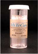 Each bottle of MicroRT™ Capillaries contains 20 – 1.5″ (37 mm) long clear polyester capillaries...